Monday, June 10, 2013

Colonial Ascidians

Kingdom Animalia
Phylum Chordata
Subphylum Tunicata
Class Ascidiacea
Colonial ascidians (AKA tunicates or sea squirts)

Our closest invertebrate relatives!  After rapid development over the course of just a few days, our sea squirts have metamorphosed and are close to maturation.  I've actually become quite fond of these little guys because they have the coolest body plan.  They have an oral and an atrial siphon that water enters and exits, respectively, through.  The water goes through mucus-covered gills into the atrium.  The mucus in the gills slits (more technically called pharyngeal slits) traps little particles from the sea water and carries them to the esophagus for digestion.  The pharyngeal slits have cilia around them to transport the mucus.

Although we do not know the species of the ascidians, we do know that they are colonial.  What does this mean?  There are  main types of ascidians: solitary and colonial.
Solitary ascidians live singularly by themselves, or form large, packed groups.  When in groups, however, they do not reproduce asexually like most other aggregating organisms.
Colonial ascidians can be divided into social or compound.  In social ascidians, individual organisms in the colony have the same body shape as if they were to stand on their own, but they are connected at the base by their stolons.  In compound ascidians, individual colony members (zooids), have their own incurrent siphons, but share one common atrial siphon with many other zooids.  So cool!
Unfortunately, it is too early in development to determine if these are social or compound ascidians.  Can't wait to find out!

Below are pictures and videos of adult sea squirts.
Fertilized 4/23/13
Images Captured 5/2/13
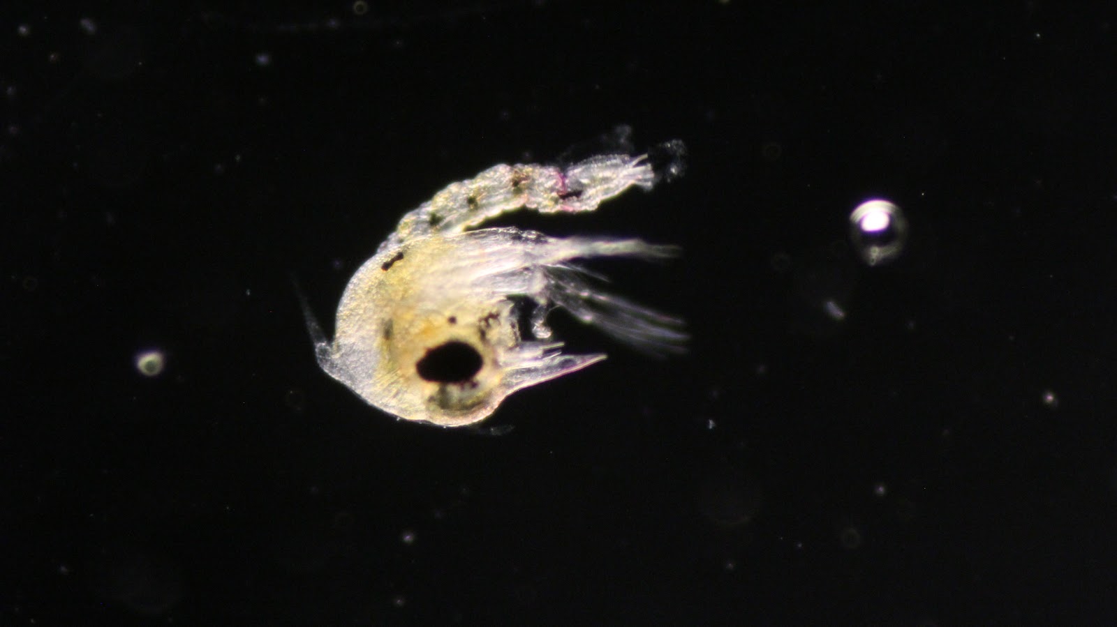
Pictures:
In this image you can clearly see the main parts of the body plan: incurrent siphon, excurrent siphon, pharyngeal slits, stomach, intestine.

Video:

Unfortunately, again, due to technical difficulties, the videos would not upload directly onto this web page, so I have attached the links to the YouTube videos.  Please read the description to get  a better idea of what you are looking at!

Gill Slits:
http://www.youtube.com/watch?v=p0UBJUsorgo
http://www.youtube.com/watch?v=le0K4IwUagU

Heart:
http://www.youtube.com/watch?v=bXD1x0mX0gU
http://www.youtube.com/watch?v=ZeITM0moiHA
-Amy Kim

Leptasterias aequalis: Brooding mother


Phylum Echinodermata
Class Asteroidea
Order Forcipulatida
Family Asteriidae
Formerly known as Leptasterias hexactis

My mother broods over things like what to make for dinner, but this tough mama broods all of her little sea star babies!  Instead of spawning into the water column like many of the species we have seen thus far, this little six armed sea star broods her babies under her oral disc from embryos to juveniles.  Brooding lasts anywhere from 6-8 weeks on average, after which the juvenile sea stars walk away on their own.  The mom does not feed during incubation - mom's are truly superheroes!  Previously, it has been observed that when the babies get dropped from the safe undersides of their mother (due to some accident), the mom swings back around and picks them up again.

Pictures:
In the first couple of pictures show the underside of the Leptasterias juvenile.  You can clearly see their six eye spots (red dots), their ambulacrum, primary podium, and mouth.


Videos:
The following videos show different behaviors and characteristics of juvenile Leptasterias and their mother.  The juveniles are now beginning to go off and explore the world of the Developmental Biology sea tables.
Note: some of the videos would not upload, so I have pasted their url.  Although this is a slight hassle, make sure you watch them because these babies are so cute.  You won't want to miss it!  Also make sure to read the descriptions of the videos when you watch!
Mother says no as she sweeps up a juvenile.
Observed: brooding mother recollects the juveniles when they are dropped

http://www.youtube.com/watch?v=GC7SzEGRg4w

http://www.youtube.com/watch?v=Ssh7ISF5zpM

http://www.youtube.com/watch?v=UKsvCVt9a00

-Amy Kim

Monday, June 3, 2013

Canis lupus familiaris

Phylum: Chordata
Order: Carnivora
Family: Canidae
Genus: Canis
Species: Canis lupus
Subspecies: Canis lupus familiaris


The domestic dog (C. lupus) is an exemplary field companion and an expert microscopy assistant. Most individuals are highly fond of echinoderms, although they can become startled around particularly active Holothuroid specimens.


They also enjoy sticks that are definitely not too big for their mouths even though they're bigger than their faces, so stop laughing at my attempts to bring one onto the trampoline even though it's so heavy I can hardly lift my head. Seriously guys.


Srsly.


---Anonymous---

Navanax inermis (?)

Two weeks ago, the class took a trip to Moss Landing to try to find intertidal worms. While wading through a tide pool in the mud, Chris Lang found a gelatinous egg mass with yellow ribbons throughout that we believe was laid by a nearby Navanax inermis (Phylum Mollusca, Class Gastropoda, Order Opistobranchia). Though this identification is only a hypothesis, this organism's developmental pattern has thus far mirrored that of my Hermissenda embryos, and the Navanax we found was the only visible Opisthobranch in the area.

It's interesting to note that these particular veligers are incredibly yolky until they hatch (more pictures to show the post-hatch transformation are coming soon!), and they have a very distinct red spot on their left-hand side (the spot will eventually darken significantly, as it has in certain individuals below).

Egg mass:


Veligers inside egg mass:


Veligers inside capsules:


Is this the culprit?


UPDATE: 
     The hatched veligers are significantly less yolky than they were when they were still encapsulated, and as mentioned above, their spots have darkened. They are just as active as the Hermissenda veligers, and are near impossible to catch resting. In the videos and pictures below, you can clearly see the eye spots at the base of the velum as well as a protrusion of either muscle or shell (it looks like shell, but it is ciliated on one side) where the shell meets the velum. Gut cilia are also active.


- Andrew Miller

Sunday, June 2, 2013

2 updates: my babies be hatching (zoea) + mystery larva from Moss Landing are settled

Hey all,

I wanted to give the class an update on the status of my crab eggs, Pachygrapsus crassipes.  I am nothing short of amazed having watched these guys develop from yolky embryos to full-fledged zoeal larvae.

Pipetting the individual eggs and larva and releasing them into a new dish with clean (2 micron filtered) sea water, rather than changing the sea water with the larvae within, is the way to go.  In this batch of eggs I am witnessing life both at its finest and most vulnerable.  Some zoea are struggling to free themselves from the strands that attach them to the eggs, while others are testing out how to move their setae to swim about in the dish.  Others were less fortunate, as the eggs simply did not continue to develop or were attacked by bacteria prematurely.  If I take away one thing from the eggs and adults of P. crassipes, it is that these are some resilient organisms!  Due to the thick membrane that encases the eggs, they can go weeks after being separated from the mother's abdomen and still manage to survive.

Notice the pointy rostrum that protrudes from the head, and the tail and setae which help for locomotion.

Beneath I am also posting a picture of the larva from our mystery egg mass from Moss Landing.  They are quite small, which makes them unideal for a close-up, in-focus picture on this camera/microscope.  Regardless, it's interesting to note how fast these eggs developed and hatched into trochophores.  Now they have graduated to veliger stage, with shell formation and all.  I am really looking forward to watching how these guys continue to develop throughout the week.


best,
 Chris

Flourescent P. giganteus

Hey all,

Here are some pictures from the first time we did immunocytochemistry. These are some of the P. giganteus on the compound microscope.


Isabel C.

Thursday, May 30, 2013

P. parvimensis Pentacula

Phylum: Holothuroideaia
Order: Aspidochirotida
Family: Stichopodidae
Genus: Parastichopus
Species: P. parvimensis

Hi all,
I discovered a parvimensis pentacula the other day! 
Here it is in all of its larval glory:

Isabel C.